nkmaxbio AAD0614说明书

nkmaxbio AAD0614说明书

Human Adiponectin/Acrp30 antibody

人脂联素/Acrp30抗体

  • Cat No.

  • AAD0614

产品类型

单克隆抗体

克隆编号5H7

UniProt编号:Q15848

NCBI登录号NP_004788

备选名称ADIPOQ、ACDC、ACRP30、APM1、GBP28、脂联素球状结构域、小鼠、脂肪细胞、C1q和含胶原蛋白结构域的蛋白质、30kDa脂肪细胞补体相关蛋白质、脂肪细胞补补体相关30kDa蛋白、ACRP40、脂肪丰富、脂联蛋白、30kda脂肪细胞补体相关蛋白质,ACRP 30、脂肪细胞C1q和含有胶原蛋白结构的蛋白质,脂肪细胞补体相关30kDa蛋白,脂肪细胞补补体相关蛋白30kDa,脂肪丰富的基因转录物1,APM1,GBP28,明胶结合蛋白

与人类反应

浓度1mg/ml(通过BCA测定)

配方含0.02%叠氮化纳、10%甘油的磷酸盐缓冲盐水(pH 7.4)液体

免疫原从大肠杆菌中纯化的重组人脂联素(15-244aa)

同种型IgG1κ

蛋白G亲和层析纯化

应用ELISA、WB、ICC/IF

用途通过ELISA、Western blot和ICC/IF分析测试抗体,以确保特异性和反应性。然而,由于应用不同,每次调查都应该用试剂滴定,以获得最佳结果。

存储可在+2C至+8C温度下存储1周。对于长期储存,等分并储存在-20℃至-80℃。避免重复冷冻和解冻循环。

    Immunflorescense (ICC/IF)

    ICC/IF analysis of Adiponectin in Hep3B cells. The cell was stained with AAD0614 (1:100). The secondary antibody (green) was used Alexa Fluor 488. DAPI was stained the cell nucleus (blue).

    Western blot analysis (WB)

    The cell lysates (10ug) were resolved by SDS-PAGE, transferred to PVDF membrane and probed with anti-human Adiponectin antibody (1:2000). Proteins were visualized using a goat anti-mouse secondary antibody conjugated to HRP and an ECL detection system.
    Lane 1. : 293T cell lysate
    Lane 2. : Adiponectin collagen domain(15-107aa) transfected 293T cell lysate
    Lane 3. : Adiponectin Full domain(15-244aa) transfected 293T cell lysate

Annexin V-PE/7-AAD细胞凋亡检测试剂盒|Annexin V-PE/7-AAD Apoptosis Detection Kit

Annexin V-PE/7-AAD细胞凋亡检测试剂盒|Annexin V-PE/7-AAD Apoptosis Detection Kit

产品说明书

FAQ

COA

已发表文献

Annexin V-PE/7-AAD细胞凋亡检测试剂盒是用红色荧光染料PE(Phycoerythrin)标记的Annexin V作为探针,来检测细胞早期凋亡的发生,可用荧光显微镜、流式细胞仪或其他荧光检测设备进行检测。

其检测原理为:在正常的活细胞中,磷脂酰丝氨酸(phosphotidylserine,PS)位于细胞膜的内侧,但在早期凋亡的细胞中,PS 从细胞膜的内侧翻转到细胞膜的表面,暴露在细胞外环境中。Annexin-(膜联蛋白-V)是一种分子量为35-36KD的Ca2+ 依赖性磷脂结合蛋白,能与PS高亲和力结合。可通过细胞外侧暴露的磷脂酰丝氨酸与凋亡早期细胞的胞膜结合。

另外,本试剂盒中提供的7-AAD可用来区分存活的早期细胞和坏死或晚期凋亡细胞。7-AAD是一种核酸染料,同PI有着相似的荧光特性,但其发射光谱较PI窄,对其他检测通道的干扰更小,在多色荧光分析中是PI的最佳替代品,可与Annexin V联合使用。该染料不能透过正常细胞或早期凋亡细胞的完整的细胞膜,但可穿透晚期凋亡细胞或者坏死细胞并与其内的DNA结合。因此将Annexin V-PE7-AAD联合使用时,7-AAD则被排除在活细胞(Annexin V-/7-AAD-)和早期凋亡细胞(Annexin V+/7-AAD-)之外,而晚期凋亡细胞和坏死细胞同时被Annexin V-PE7-AAD结合染色呈现双阳性(Annexin V+/7-AAD+)。


产品组分

编号

组分名称

产品编号/规格

40310ES20

20T

40310ES50

50T

40310ES60

100T

40310-A

重组人Annexin V/PE*(rh Annexin V/PE)

100 µL

250 µL

500 µL

40310-B

7-AAD Viability Staining Solution(20µg/mL)

200 µL

500 µL

1.0 mL

40310-C

4×Binding Buffer**

4 mL

10 mL

20 mL

*来源于大肠杆菌(E.coli),分子量为35.8 KDa,纯度>98%(SDS-PAGE & HPLC)


运输与保存方法

冰袋(wet ice)运输。本试剂盒所有组分均在4 保存,勿冰冻Annexin V-PE、7-AAD需避光保存。一年有效。


注意事项

1)试剂在开盖前请短暂离心,将盖内壁上的液体甩至管底,避免开盖时液体洒落。

2)由于细胞凋亡是一个快速的过程,建议样品在染色后1小时之内进行分析。

3)对于贴壁细胞,消化是一个关键步骤。贴壁细胞诱导细胞凋亡时如有漂浮细胞,需收集漂浮细胞和贴壁细胞后合并染色。处理贴壁细胞时要小心操作,尽量避免人为的损伤细胞。胰酶消化时间过短,细胞需要用力吹打才能脱落,容易造成细胞膜的损伤,7-AAD摄入过多;消化时间过长,细胞膜同样易造成损伤,甚至会影响细胞膜上磷脂酰丝氨酸与Annexin V-PE的结合。消化时将胰酶铺满孔板底后,轻摇时胰酶与细胞充分接触,然后倒掉大部分胰酶,利用剩余的少量胰酶再消化一段时间,待细胞间空隙增大,瓶底呈花斑状即可终止。在消化液中尽量不用EDTA,EDTA会影响Annexin V与PS的结合。

4)如果样品来源于血液,请务必除去血液中的血小板。因为血小板含有PS,能与Annexin V结合,从而干扰实验结果。可以使用含有EDTA的缓冲剂并低速离心(200×g)洗去血小板,然后用Annexin V结合缓冲液清洗细胞,以避免残留的EDTA会螯合Ca2+

57-AAD为潜在致癌物,操作时请采取防护措施,穿防护服、戴手套等

6Annexin V-PE7-AAD是光敏物质,在操作时请注意避光。

7)本产品仅作科研用途!


使用说明

1. 样品染色

1) 悬浮细胞:300 g,4 ℃离心5 min收集细胞。

贴壁细胞:用不含EDTA的胰酶消化后,300g,4 ℃离心5 min收集细胞。胰酶消化时间不宜过长,以防引起假阳性。

2)配制1×Binding Buffer:用去离子水4倍稀释4×Binding Buffer4 mL 结合缓冲液+12 mL 去离子水)

3)4 ℃预冷的PBS洗涤细胞2次,每次均需300 g,4 ℃离心5 min。

4)加入250 μL 1×Binding Buffer重悬细胞,调节其浓度为1×10⁶细胞/mL

5)取100 μL细胞悬液于5 mL流式管中,加入5 μL Annexin V/PE10 μL 7-AAD,轻轻混匀。

6)避光、室温反应15 min。

7)加入400 μL 1×Binding Buffer,混匀,样品在1小时内检测。

【注】:为了避免洗涤细胞时损失细胞,在吸液时可以用大的Tip头套上小的Tip头吸液。

2. 观察检测

A、流式细胞仪分析

1)用流式细胞仪检测,Annexin V-PE的激发波长Ex=488nm;发射波长Em=578 nm,发出橙红色荧光,建议使用FL2通道检测;7-AAD的激发波长Ex=546 nm;发射波长Em=647 nm,发出红色荧光,建议使用FL3通道检测CellQuest等软件进行分析,绘制双色散点图(two-color dot plot),PE为横坐标,7-AAD为纵坐标。每个样采集10,000 events。典型的实验中,细胞可以分成三个亚群,活细胞仅很低强度的背景荧光,早期凋亡细胞仅较强的橙红色荧光,晚期凋亡细胞呈橙红和红色荧光双重染色。

2)荧光补偿调节:使用未经凋亡诱导处理的正常细胞,作为对照进行荧光补偿调节去除光谱重叠和设定十字门的位置。

B、荧光显微镜观察

1)滴一滴上述染色后的细胞悬液于载玻片上,并用盖玻片盖上细胞;

2)对于贴壁细胞来说,也可直接用盖玻片来培养细胞并诱导细胞凋亡;

a 将细胞于盖玻片上生长,用适当的凋亡诱导剂诱导细胞凋亡,并设立阴性对照组;

b 用PBS洗涤细胞两次;

c 在500 μL的Binding Buffer 中加入1 μL Annexin V-PE,5 μL 7-AAD染液混匀;

d 将上述溶液滴加于盖玻片表面,使长有细胞的盖玻片表面均匀覆盖;

e 室温避光孵育5 min。

3)将盖玻片倒置于载玻片上,在荧光显微镜下用双色滤光片观察。滤光片(罗丹明)观察Annexin V-PE 荧光信号呈橙红色;用激发波长546 nm,发射波长647 nm,观察7-AAD荧光信号呈红色。


HB210802


Q:这两个试剂用什么通道检测?仪器是贝克曼的。

A: Annexin V-PE 的激发波长Ex=488nm;发射波长Em=578 nm,发出橙红色荧光,建议使用 FL2 通道检测;7-AAD 的激发波长 Ex=546 nm;发射波长 Em=647 nm,发出红色荧光, 建议使用 FL3 通道检测。

Q: Annexin V-PE 染色失败或者阳性率偏低。

A:首先要确定实验中使用的诱导剂是否能产生凋亡。可通过设定确切凋亡诱导效果的阳性药物对照来排除这一情况。Annexin V-PE 染色失败,最常见的实验操作原因是贴壁细胞消化不当。Annexin V 跟 PS 的结合需要 Ca2+,Binding Buffer 中含有 2.5 mM 的 Ca2+,含 EDTA 的胰酶消化会影响染色,建议使用无EDTA 的胰酶。若使用含EDTA 的胰酶,必须通过洗涤步骤彻底去除EDTA。用 PBS 洗涤细胞沉淀后,应尽量去除残余液体。残留PBS 中的磷酸根,会形成磷酸钙沉淀。Binding Buffer 的瓶盖要紧闭,防止空气中的 CO2 进入后形成碳酸钙沉淀,减少游离Ca2+,导致实验失败。接触其他缓冲液如吸取PBS 后不更换枪头也会导致游离的 Ca2+减少。一些细胞其细胞膜上 PS 的密度较低,染色效果很差,此时建议更换细胞株或者采用 TUNEL 试剂盒检测凋亡。如果是贴壁细胞,药物诱导后漂浮的细胞也要收集,这部分细胞往往是凋亡阳性的细胞,丢弃会造成阳性结果偏低。

Q: 假阳性

A: 实验中发现未经诱导凋亡的对照细胞经染色后Annexin V-PE/7-AAD 双阳性比例过高,造成这种结果的原因可能是细胞本身活力低。建议用台盼蓝染色计算细胞活力,阴性对照台盼蓝拒染的细胞应大于 95%。若细胞活力低,建议重新复苏细胞,通常刚复苏的细胞至少应传代 2– 3 次以后才能进行实验。另一可能是细胞操作不当引起,操作过程中吹打细胞过于剧烈,贴壁细胞消化过程中消化时间过长,均可能导致细胞膜破坏,出现假阳性。此外,一般情况下诱导几个小时就可以出现早期凋亡,因此通常不需要处理大于 48 h 以上;诱导时间过长会使营养物质耗尽,导致细胞状态差,假阳性结果偏高。

[1] Shi J, Ju R, Gao H, Huang Y, Guo L, Zhang D. Targeting glutamine utilization to block metabolic adaptation of tumor cells under the stress of carboxyamidotriazole-induced nutrients unavailability. Acta Pharm Sin B. 2022;12(2):759-773. doi:10.1016/j.apsb.2021.07.008(IF:11.614)
[2] Liang C, Zou T, Zhang M, et al. MicroRNA-146a switches microglial phenotypes to resist the pathological processes and cognitive degradation of Alzheimer's disease. Theranostics. 2021;11(9):4103-4121. Published 2021 Feb 19. doi:10.7150/thno.53418(IF:11.556)
[3] Li J, Li J, Yao Y, et al. Biodegradable electrospun nanofibrous platform integrating antiplatelet therapy-chemotherapy for preventing postoperative tumor recurrence and metastasis. Theranostics. 2022;12(7):3503-3517. Published 2022 Apr 24. doi:10.7150/thno.69795(IF:11.556)
[4] Yang Y, Qiao X, Huang R, et al. E-jet 3D printed drug delivery implants to inhibit growth and metastasis of orthotopic breast cancer. Biomaterials. 2020;230:119618. doi:10.1016/j.biomaterials.2019.119618(IF:10.273)
[5] Wang D, Zhang L, Hu A, et al. Loss of 4.1N in epithelial ovarian cancer results in EMT and matrix-detached cell death resistance. Protein Cell. 2021;12(2):107-127. doi:10.1007/s13238-020-00723-9(IF:10.164)
[6] Fan Z, Chen X, Liu L, et al. Association of the Polymorphism rs13259960 in SLEAR With Predisposition to Systemic Lupus Erythematosus. Arthritis Rheumatol. 2020;72(6):985-996. doi:10.1002/art.41200(IF:9.586)
[7] Xu Y, Hu Y, Xu T, et al. RNF8-mediated regulation of Akt promotes lung cancer cell survival and resistance to DNA damage. Cell Rep. 2021;37(3):109854. doi:10.1016/j.celrep.2021.109854(IF:9.423)
[8] Zhu C, Zhang L, Zhao S, et al. UPF1 promotes chemoresistance to oxaliplatin through regulation of TOP2A activity and maintenance of stemness in colorectal cancer. Cell Death Dis. 2021;12(6):519. Published 2021 May 21. doi:10.1038/s41419-021-03798-2(IF:8.469)
[9] Yang YL, Zhang Y, Li DD, et al. RNF144A functions as a tumor suppressor in breast cancer through ubiquitin ligase activity-dependent regulation of stability and oncogenic functions of HSPA2. Cell Death Differ. 2020;27(3):1105-1118. doi:10.1038/s41418-019-0400-z(IF:8.086)
[10] Yuan S, Zheng P, Sun X, et al. Hsa_Circ_0001860 Promotes Smad7 to Enhance MPA Resistance in Endometrial Cancer via miR-520h. Front Cell Dev Biol. 2021;9:738189. Published 2021 Nov 29. doi:10.3389/fcell.2021.738189(IF:6.684)
[11] Lian Y, Hao H, Xu J, Bo T, Wang W. Histone Chaperone Nrp1 Mutation Affects the Acetylation of H3K56 in Tetrahymena thermophila. Cells. 2022;11(3):408. Published 2022 Jan 25. doi:10.3390/cells11030408(IF:6.600)
[12] Cai B, Hou M, Zhang S, et al. Dual Targeting of Endoplasmic Reticulum by Redox-Deubiquitination Regulation for Cancer Therapy. Int J Nanomedicine. 2021;16:5193-5209. Published 2021 Jul 30. doi:10.2147/IJN.S321612(IF:6.400)
[13] Liu G, Liu B, Liu X, et al. ARID1B/SUB1-activated lncRNA HOXA-AS2 drives the malignant behaviour of hepatoblastoma through regulation of HOXA3. J Cell Mol Med. 2021;25(7):3524-3536. doi:10.1111/jcmm.16435(IF:5.310)
[14] He J, Zhao T, Liu L, et al. The -172 A-to-G variation in ADAM17 gene promoter region affects EGR1/ADAM17 pathway and confers susceptibility to septic mortality with sepsis-3.0 criteria. Int Immunopharmacol. 2022;102:108385. doi:10.1016/j.intimp.2021.108385(IF:4.932)
[15] Yin H, Wang H, Wang M, et al. CircTCF25 serves as a sponge for miR-206 to support proliferation, migration, and invasion of glioma via the Jak2/p-Stat3/CypB axis. Mol Carcinog. 2022;61(6):558-571. doi:10.1002/mc.23402(IF:4.784)
[16] Lin L, Li H, Shi D, et al. Depletion of C12orf48 inhibits gastric cancer growth and metastasis via up-regulating Poly r(C)-Binding Protein (PCBP) 1. BMC Cancer. 2022;22(1):123. Published 2022 Jan 31. doi:10.1186/s12885-022-09220-0(IF:4.430)
[17] Wang P, Zhang C, Li J, et al. Adipose-derived mesenchymal stromal cells improve hemodynamic function in pulmonary arterial hypertension: identification of microRNAs implicated in modulating endothelial function. Cytotherapy. 2019;21(4):416-427. doi:10.1016/j.jcyt.2019.02.011(IF:4.297)
[18] Cao K, Chen Y, Zhao S, et al. Sirt3 Promoted DNA Damage Repair and Radioresistance Through ATM-Chk2 in Non-small Cell Lung Cancer Cells. J Cancer. 2021;12(18):5464-5472. Published 2021 Jul 13. doi:10.7150/jca.53173(IF:4.207)
[19] Cao Y, Chu C, Li X, Gu S, Zou Q, Jin Y. RNA-binding protein QKI suppresses breast cancer via RASA1/MAPK signaling pathway. Ann Transl Med. 2021;9(2):104. doi:10.21037/atm-20-4859(IF:3.932)
[20] Jiang ZQ, Li MH, Qin YM, Jiang HY, Zhang X, Wu MH. Luteolin Inhibits Tumorigenesis and Induces Apoptosis of Non-Small Cell Lung Cancer Cells via Regulation of MicroRNA-34a-5p. Int J Mol Sci. 2018;19(2):447. Published 2018 Feb 2. doi:10.3390/ijms19020447(IF:3.687)
[21] Qin Y, Liang Y, Jiang G, Peng Y, Feng W. ACY-1215 suppresses the proliferation and induces apoptosis of chronic myeloid leukemia cells via the ROS/PTEN/Akt pathway [published online ahead of print, 2022 Jun 8]. Cell Stress Chaperones. 2022;10.1007/s12192-022-01280-2. doi:10.1007/s12192-022-01280-2(IF:3.667)
[22] Wu S, Luo C, Li F, Hameed NUF, Jin Q, Zhang J. Silencing expression of PHF14 in glioblastoma promotes apoptosis, mitigates proliferation and invasiveness via Wnt signal pathway. Cancer Cell Int. 2019;19:314. Published 2019 Nov 27. doi:10.1186/s12935-019-1040-6(IF:3.439)
[23] Dai H, Wang J, Huang Z, et al. LncRNA OIP5-AS1 Promotes the Autophagy-Related Imatinib Resistance in Chronic Myeloid Leukemia Cells by Regulating miR-30e-5p/ATG12 Axis. Technol Cancer Res Treat. 2021;20:15330338211052150. doi:10.1177/15330338211052150(IF:3.399)
[24] Wu S, Luo C, Hameed NUF, Wang Y, Zhuang D. UCP2 silencing in glioblastoma reduces cell proliferation and invasiveness by inhibiting p38 MAPK pathway. Exp Cell Res. 2020;394(1):112110. doi:10.1016/j.yexcr.2020.112110(IF:3.383)
[25] Chen A, Fang D, Ren Y, Wang Z. Matrine protects colon mucosal epithelial cells against inflammation and apoptosis via the Janus kinase 2 /signal transducer and activator of transcription 3 pathway. Bioengineered. 2022;13(3):6490-6499. doi:10.1080/21655979.2022.2031676(IF:3.269)
[26] Jiang K, Zou H. microRNA-20b-5p overexpression combing Pembrolizumab potentiates cancer cells to radiation therapy via repressing programmed death-ligand 1. Bioengineered. 2022;13(1):917-929. doi:10.1080/21655979.2021.2014617(IF:3.269)
[27] Liu S, Xin Y, Shi J, et al. ML365 inhibits lipopolysaccharide-induced inflammatory responses via the NF-κB signaling pathway. Immunobiology. 2022;227(3):152208. doi:10.1016/j.imbio.2022.152208(IF:3.144)
[28] Wang Z, Fu L, Zhang J, et al. A comprehensive analysis of potential gastric cancer prognostic biomarker ITGBL1 associated with immune infiltration and epithelial-mesenchymal transition. Biomed Eng Online. 2022;21(1):30. Published 2022 May 20. doi:10.1186/s12938-022-00998-5(IF:2.819)
[29] Sun Y, Tian X, Wu H, et al. H2O2 signaling modulates Glycoprotein-1 induced programmed cell death in tobacco suspension cells. Pestic Biochem Physiol. 2021;171:104697. doi:10.1016/j.pestbp.2020.104697(IF:2.751)
[30] Zhao L, Zhou R, Wang Q, Cheng Y, Gao M, Huang C. MicroRNA-320c inhibits articular chondrocytes proliferation and induces apoptosis by targeting mitogen-activated protein kinase 1 (MAPK1). Int J Rheum Dis. 2021;24(3):402-410. doi:10.1111/1756-185X.14053(IF:2.454)
[31] Yao T, Wang L, Ding ZF, Yin ZS. hsa_circ_0058122 knockdown prevents steroid-induced osteonecrosis of the femoral head by inhibiting human umbilical vein endothelial cells apoptosis via the miR-7974/IGFBP5 axis. J Clin Lab Anal. 2022;36(4):e24134. doi:10.1002/jcla.24134(IF:2.352)
[32] Zhong W, Zhao Y, Tian Y, Chen M, Lai X. The protective effects of HGF against apoptosis in vascular endothelial cells caused by peripheral vascular injury. Acta Biochim Biophys Sin (Shanghai). 2018;50(7):701-708. doi:10.1093/abbs/gmy048(IF:2.224)
[33] Zhang Z, Yi J, Xie B, et al. Parkin, as a Regulator, Participates in Arsenic Trioxide-Triggered Mitophagy in HeLa Cells. Curr Issues Mol Biol. 2022;44(6):2759-2771. Published 2022 Jun 20. doi:10.3390/cimb44060189(IF:2.081)
[34] Fan Y, Tan D, Zhang X, et al. Nuclear Factor-κB Pathway Mediates the Molecular Pathogenesis of LMNA-Related Muscular Dystrophies. Biochem Genet. 2020;58(6):966-980. doi:10.1007/s10528-020-09989-4(IF:2.027)
[35] Li M, Min W, Wang J, et al. Effects of mevalonate kinase interference on cell differentiation, apoptosis, prenylation and geranylgeranylation of human keratinocytes are attenuated by farnesyl pyrophosphate or geranylgeranyl pyrophosphate. Exp Ther Med. 2020;19(4):2861-2870. doi:10.3892/etm.2020.8569(IF:1.785)
[36] Zhao L. Protective effects of trimetazidine and coenzyme Q10 on cisplatin-induced cardiotoxicity by alleviating oxidative stress and mitochondrial dysfunction. Anatol J Cardiol. 2019;22(5):232-239. doi:10.14744/AnatolJCardiol.2019.83710(IF:1.112)

7-AAD 7-氨基放线菌素D 核酸染料|7-AAD

7-AAD 7-氨基放线菌素D 核酸染料|7-AAD

产品说明书

FAQ

COA

已发表文献

产品信息


产品名称        

产品编号

规格

储存

价格(元)

7-AAD

7-氨基放线菌素D

40722ES03

1mg

-20℃

1438.00

产品描述
 

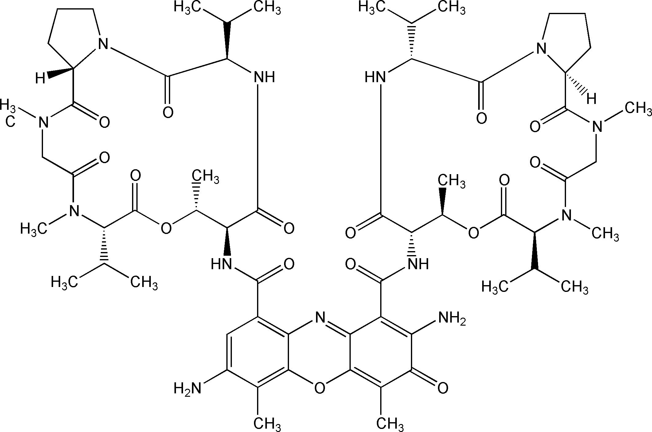
7-Aminoactinomycin D,简称7-AAD,是一种核酸染料,与DNA结合后可发出强烈的荧光,其荧光特性与PI相似,可被488nm氩离子激光激发,但其发射光谱较PI窄,且发射波长更长,对其他检测通道的干扰更小,在多色荧光分析中是PI的最佳替代品,可与多种488激发光激发的荧光染料联合使用,如FITC,PE等。

另外,7-AAD是一种非渗透性荧光染料。该染料不能透过活细胞的细胞膜,但可穿透膜损伤细胞如晚期凋亡细胞或者坏死细胞的细胞膜并与其内的DNA结合,可用来区分存活的早期细胞和坏死或晚期凋亡细胞,广泛用于流式细胞仪。

产品性质
 

英文别名(English Synonym)

7-Aminoactinomycin D; Actinomycin   D, 7-amino-

CAS号(CAS NO.)

7240-37-1

分子式(Formula)

C62H87N13O16

分子量(Molecular   Weight)

1270.43

外观(Appearance)

Solid

Ex/Em

546 nm/647 nm

溶解性(Solubility)

溶于水和DMSO.

结构式(Structure)

7-AAD 7-氨基放线菌素D 核酸染料|7-AAD

运输和保存方法


室温运输,-20℃干燥避光保存,有效期一年。
 

注意事项


1)7-AAD为潜在致癌物质,操作时请穿戴手套、眼罩以及其他防护措施,以避免接触皮肤。
2)玻璃器皿上残留的去垢剂也会影响染色的效果,导致细胞存在或不存在都会出现强荧光。玻璃器皿的清洗应用去垢剂,然后用自来水冲洗数遍后,用去离子水或蒸馏水漂洗。

3)为了您的安全和健康,请穿实验服并戴一次性手套操作。
4) 本产品仅作科研用途!


使用方法
 

注:该操作说明适用于大多数细胞,但不同的细胞类型、细胞密度、使用的培养基以及其他一些因素都有可能影响染色效果,本说明仅供参考。

1)储存液的制备:取适量DMSO加入到7-AAD配制成1-10 mM,该储存液可分装于-20℃稳定保存6个月。

2)根据自身样本选择合适的步骤固定细胞。7-AAD染色一般在其它染色完成后再进行,且仅仅染色死细胞。

3)离心收集细胞,并用合适的的缓冲盐溶液或者培养基(pH=7.4)重悬细胞。

注:贴壁细胞可在盖玻片或者培养板上进行原位染色。

4)加入适量7-AAD染色液,推荐该染料的工作浓度为0.5-5 µM,孵育15-60 min。

注:首次实验建议在推荐的工作浓度范围内设置浓度梯度检测,从而摸索最佳的染色浓度。

5)根据其激发发射波长(Ex/Em=545 nm/650 nm)在流式细胞仪下(FL3通道)检测荧光强度。


相关产品
 

货号 产品名称 规格

40745ES64

7-AAD Viability Staining Solution

7-AAD细胞活力染色液

150 T

40309ES20

Annexin V-APC/7-AAD Apoptosis Detection Kit 

Annexin V-APC/7-AAD细胞凋亡检测试剂盒

20 T

40309ES50

50 T

40309ES60

100 T

40310ES20

Annexin V-PE/7-AAD Apoptosis Detection Kit

Annexin V-PE/7-AAD细胞凋亡检测试剂盒

20 T

40310ES50

50 T

40310ES60

100 T


7-AAD 7-氨基放线菌素D 核酸染料|7-AAD

暂无内容

[1] Zhang C, Lei JL, Zhang H, et al. Calyxin Y sensitizes cisplatin-sensitive and resistant hepatocellular carcinoma cells to cisplatin through apoptotic and autophagic cell death via SCF βTrCP-mediated eEF2K degradation. Oncotarget. 2017;8(41):70595-70616. Published 2017 Aug 3. doi:10.18632/oncotarget.19883(IF:5.168)
[2] Ye M, Ma J, Liu B, Liu X, Ma D, Dong K. Linc01105 acts as an oncogene in the development of neuroblastoma [published correction appears in Oncol Rep. 2021 Sep;46(3):]. Oncol Rep. 2019;42(4):1527-1538. doi:10.3892/or.2019.7257(IF:3.041)
[3] Yu T, Xia Q, Gong T, Wang J, Zhong D. Molecular mechanism of acquired drug resistance in the EGFR-TKI resistant cell line HCC827-TR. Thorac Cancer. 2020;11(5):1129-1138. doi:10.1111/1759-7714.13342(IF:2.610)

7-AAD细胞活力染色液 7-AAD核酸染料|7-AAD Viability Staining Solution

7-AAD细胞活力染色液 7-AAD核酸染料|7-AAD Viability Staining Solution

产品说明书

FAQ

COA

已发表文献

产品描述

7-Aminoactinomycin D,简称7-AAD,是一种核酸染料,与DNA结合后可发出强烈的荧光,其荧光特性与PI相似,可被488 nm氩离子激光激发,但其发射光谱较PI窄,且发射波长更长,对其他检测通道的干扰更小,在多色荧光分析中是PI的最佳替代品,可与多种488激发光激发的荧光染料联合使用,如FITC,PE等。

另外,7-AAD是一种非渗透性荧光染料。该染料不能透过活细胞的细胞膜,但可穿透膜损伤细胞如晚期凋亡细胞或者坏死细胞的细胞膜并与其内的DNA结合,可用来区分存活的早期细胞和坏死或晚期凋亡细胞,广泛用于流式细胞术检测。

本品为7-AAD的即用型染色液,可直接用于细胞染色,其最大激发波长为546nm,最大发射波长为647 nm,可在流式细胞仪下(FL3通道)检测荧光强度。若每次(含1×10⁵个细胞)用量10 µL,本品可供检测150次。

运输和保存方法

冰袋运输,4℃干燥避光保存,有效期六个月。

注意事项

1)7-AAD为潜在致癌物质,操作时请穿戴手套、眼罩以及其他防护措施,以避免接触皮肤。

2)玻璃器皿上残留的去垢剂也会影响染色的效果,导致细胞存在或不存在都会出现强荧光。玻璃器皿的清洗应用去垢剂,然后用自来水冲洗数遍后,用去离子水或蒸馏水漂洗。

3)为了您的安全和健康,请穿实验服并戴一次性手套操作。

4)本产品仅作科研用途!

使用方法

:7-AAD染色一般在其它染色完成后再进行,且仅仅染色死细胞。
样品在完成其他染色后,取0.5 mL细胞悬液(~1×10⁵个细胞),加入约10 µL 7-AAD细胞活力染色剂,根据其激发发射波长(Ex/Em=545 nm/650 nm)在流式细胞仪下(FL3通道)检测荧光强度。


相关产品

40722ES03

7-AAD 7-氨基放线菌素D

1mg

40310ES20

Annexin V-PE/7-AAD细胞凋亡检测试剂盒 Annexin V-PE/7-AAD Apoptosis Detection Kit

20 T

40310ES50

Annexin V-PE/7-AAD细胞凋亡检测试剂盒 Annexin V-PE/7-AAD Apoptosis Detection Kit

50 T

40310ES60

Annexin V-PE/7-AAD细胞凋亡检测试剂盒 Annexin V-PE/7-AAD Apoptosis Detection Kit

100 T

 
HB211104

Q:7-AAD的即用型染色液试剂浓度是多少?

A: 10 μg/ml

[1] Zhou S, Zang S, Hu Y, et al. Transcriptome-scale spatial gene expression in rat arcuate nucleus during puberty. Cell Biosci. 2022;12(1):8. Published 2022 Jan 21. doi:10.1186/s13578-022-00745-2(IF:7.133)
[2] Li Y, Dong Y, Ran Y, et al. Three-dimensional cultured mesenchymal stem cells enhance repair of ischemic stroke through inhibition of microglia. Stem Cell Res Ther. 2021;12(1):358. Published 2021 Jun 21. doi:10.1186/s13287-021-02416-4(IF:6.832)