
嘌呤霉素Puromycin 嘌呤霉素盐酸盐 氨基糖苷类抗生素|CAS 58-58-2
产品说明书
FAQ
COA
已发表文献
嘌呤霉素(Puromycin)是由白黑链霉菌(Streptomyces alboniger)发酵代谢产生的一种氨基糖苷类抗生素,通过抑制蛋白质合成而杀死革兰氏阳性菌,各种动物和昆虫细胞。某种特殊情况下有效作用大肠杆菌。作用机制在于嘌呤霉素是氨酰-tRNA分子3´末端的类似物,能够与核糖体的A位点结合并掺入到延伸的肽链中。嘌呤霉素同A位点结合后,不会参与随后的任何反应,从而导致蛋白质合成的提前终止并释放出C-末端含有嘌呤霉素的不成熟多肽。
嘌呤霉素产生菌Streptomyces alboniger内发现的pac基因编码嘌呤霉素N-乙酰转移酶(PAC),赋予机体对嘌呤霉素产生抗性。这一特性如今普遍应用于筛选特定携带pac基因质粒的哺乳动物稳定转染细胞株。
嘌呤霉素在细胞稳转株筛选中的普遍应用与慢病毒载体的特性有关,现在商业化的慢病毒载体多数都携带pac基因。在某些特定情况下,嘌呤霉素亦可以用来筛选转化携带pac基因质粒的大肠杆菌菌株。
产品信息
|
货号 |
60210JP25/60210JP60/60210JP72/60210JP76/60210JP80 |
|
规格 |
25 mg/100 mg/250 mg/500 mg/1 g |
|
CAS号(CAS NO.) |
58-58-2 |
|
分子式(Molecular Fomular) |
C22H29N7O5·2HCl |
|
分子量(Molecular Weight) |
544.43 g/mol |
|
纯度(Purity) |
≥98% |
|
外观(Appearance) |
白色至米白色粉末 |
|
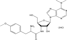
结构(Structure) |
|

储存条件
-25~-15℃保存,有效期2年。
使用说明
1. 建议使用浓度
哺乳动物细胞:1-10 μg/mL,最佳浓度需要杀灭曲线来确定。
大肠杆菌:LB琼脂培养基筛选稳定转化pac基因的大肠杆菌,使用浓度为125 μg/mL。
【注】:使用嘌呤霉素筛选大肠杆菌稳转株需要精确的pH值调节,而且受宿主细胞本身的影响。
2.溶解方法
用蒸馏水溶解嘌呤霉素配制成50 mg/mL的母液,经0.22 μm滤膜过滤除菌后分装于-25~-15℃冻存;也可溶于甲醇,配制成10 mg/mL的储存液。
3. 嘌呤霉素杀灭曲线的确定(以shRNA转染或者慢病毒转导为例)
嘌呤霉素有效筛选浓度跟细胞类型、生长状态、细胞密度、细胞代谢情况及细胞所处细胞周期位置等有关。为了筛选到稳定表达的shRNA细胞株,确定杀死未转染/转导细胞的最低浓度嘌呤霉素至关重要。建议初次做实验的客户一定要建立适合自身实验体系的杀死曲线(kill curve)。
1)Day 1:24孔板内以5~8×10⁴ cells/孔的密度铺板,铺足够量的孔以进行后续的梯度实验。37℃细胞孵育过夜。
2)Day 2:①准备筛选培养基:含不同浓度嘌呤霉素的新鲜培养基(如0-15 μg/mL,至少5个梯度);②往孵育过夜后的细胞内更换新鲜配制的筛选培养基;之后37℃孵育细胞。
3)Day 4:更换新鲜的筛选培养基,并观察细胞存活率。
4)根据细胞的生长状态,约2-3天更换新鲜的筛选培养基。
5)每日监测细胞,观察存活细胞率,从而确定抗生素筛选开始4-6天内有效杀死非转染或所有非转导细胞的药物最低浓度。
4. 哺乳动物稳定转染细胞株的筛选
等转染含有pac基因的质粒后,细胞在含有嘌呤霉素的培养基中增殖,以筛选出稳定转染子。
1)细胞转染48 h后,将细胞(原样或稀释)置于含有适当浓度嘌呤霉素的新鲜培养基中培养。
【注】:当细胞处于分裂活跃期时,抗生素作用最明显。细胞过于密集,抗生素产生的效力会明显下降。最好进行细胞分盘使其密度不超过25%。
2)每隔2-3天,移除和更换含有嘌呤霉素的培养基。
3)筛选7天后评估细胞形成的病灶。病灶可能需要额外的一周或者更多时间,这依赖于宿主细胞系和转染筛选效率。
【注】:每日进行细胞生长状态的观察。嘌呤霉素的筛选至少需要48 h,有效浓度嘌呤霉素的筛选周期一般在3-10天。
4)转移和放置5-10个抗性克隆到一个35 mm的培养皿中,用选择培养基继续培养7天。此次富集培养是为日后的细胞毒性实验做准备。
注意事项
1.嘌呤霉素为有毒化合物,操作时请小心拿放。
2.为了您的安全和健康,请穿实验服并戴一次性手套操作。
3.本产品仅用于科研用途,禁止用于人身上。
相关产品推荐:
|
产品名称 |
产品编号 |
|
Carbenicillin, Disodium Salt 羧苄青霉素钠 |
60202JP |
|
Ampicillin, Sodium Salt 氨苄青霉素钠 |
60203 |
|
Doxycycline hyclate 盐酸强力霉素 |
60204JP |
|
Chloramphenicol, USP Grade 氯霉素,USP级 |
60205JP |
|
Kanamycin Sulfate 硫酸卡那霉素 |
60206JP |
|
Puromycin Dihydrochloride嘌呤霉素盐酸盐 |
60210JP |
|
Tetracyclin HCl 四环素盐酸盐(USP) |
60212JP |
|
Vancomycin Hydrochloride 盐酸万古霉素 |
60213JP |
|
Gentamycin Sulfate Salt 硫酸庆大霉素 |
60214JP |
|
Spectinomycin Hydrochloride 盐酸壮观霉素 |
60215JP |
|
盐酸博莱霉素 |
60216JP |
|
Phleomycin (20mg/ml in solution) 腐草霉素 |
60217JP |
|
Blasticidin S 杀稻瘟菌素S (灭瘟素) |
60218JP |
|
Nystatin 制真菌素 |
60219JP |
|
G418 Sulfate (Geneticin) 遗传霉素 |
60220JP |
|
Hygromycin B 潮霉素B |
60225JP |
|
Erythromycin 红霉素 |
60228JP |
|
Timentin 特美汀 |
60230JP |
|
Aureobasidin A (AbA)金担子素A |
60231JP |
|
Polymyxin B Sulfate 多粘菌素B硫酸盐 |
60242JP |
嘌呤霉素(Puromycin)是由白黑链霉菌(Streptomyces alboniger)发酵代谢产生的一种氨基糖苷类抗生素,通过抑制蛋白质合成而杀死革兰氏阳性菌,各种动物和昆虫细胞。某种特殊情况下有效作用大肠杆菌。作用机制在于嘌呤霉素是氨酰-tRNA分子3´末端的类似物,能够与核糖体的A位点结合并掺入到延伸的肽链中。嘌呤霉素同A位点结合后,不会参与随后的任何反应,从而导致蛋白质合成的提前终止并释放出C-末端含有嘌呤霉素的不成熟多肽。
嘌呤霉素产生菌Streptomyces alboniger内发现的pac基因编码嘌呤霉素N-乙酰转移酶(PAC),赋予机体对嘌呤霉素产生抗性。这一特性如今普遍应用于筛选特定携带pac基因质粒的哺乳动物稳定转染细胞株。
嘌呤霉素在细胞稳转株筛选中的普遍应用与慢病毒载体的特性有关,现在商业化的慢病毒载体多数都携带pac基因。在某些特定情况下,嘌呤霉素亦可以用来筛选转化携带pac基因质粒的大肠杆菌菌株。
产品信息
|
货号 |
60210JP25/60210JP60/60210JP72/60210JP76/60210JP80 |
|
规格 |
25 mg/100 mg/250 mg/500 mg/1 g |
|
CAS号(CAS NO.) |
58-58-2 |
|
分子式(Molecular Fomular) |
C22H29N7O5·2HCl |
|
分子量(Molecular Weight) |
544.43 g/mol |
|
纯度(Purity) |
≥98% |
|
外观(Appearance) |
白色至米白色粉末 |
|
结构(Structure) |
|

储存条件
-25~-15℃保存,有效期2年。
使用说明
1. 建议使用浓度
哺乳动物细胞:1-10 μg/mL,最佳浓度需要杀灭曲线来确定。
大肠杆菌:LB琼脂培养基筛选稳定转化pac基因的大肠杆菌,使用浓度为125 μg/mL。
【注】:使用嘌呤霉素筛选大肠杆菌稳转株需要精确的pH值调节,而且受宿主细胞本身的影响。
2.溶解方法
用蒸馏水溶解嘌呤霉素配制成50 mg/mL的母液,经0.22 μm滤膜过滤除菌后分装于-25~-15℃冻存;也可溶于甲醇,配制成10 mg/mL的储存液。
3. 嘌呤霉素杀灭曲线的确定(以shRNA转染或者慢病毒转导为例)
嘌呤霉素有效筛选浓度跟细胞类型、生长状态、细胞密度、细胞代谢情况及细胞所处细胞周期位置等有关。为了筛选到稳定表达的shRNA细胞株,确定杀死未转染/转导细胞的最低浓度嘌呤霉素至关重要。建议初次做实验的客户一定要建立适合自身实验体系的杀死曲线(kill curve)。
1)Day 1:24孔板内以5~8×10⁴ cells/孔的密度铺板,铺足够量的孔以进行后续的梯度实验。37℃细胞孵育过夜。
2)Day 2:①准备筛选培养基:含不同浓度嘌呤霉素的新鲜培养基(如0-15 μg/mL,至少5个梯度);②往孵育过夜后的细胞内更换新鲜配制的筛选培养基;之后37℃孵育细胞。
3)Day 4:更换新鲜的筛选培养基,并观察细胞存活率。
4)根据细胞的生长状态,约2-3天更换新鲜的筛选培养基。
5)每日监测细胞,观察存活细胞率,从而确定抗生素筛选开始4-6天内有效杀死非转染或所有非转导细胞的药物最低浓度。
4. 哺乳动物稳定转染细胞株的筛选
等转染含有pac基因的质粒后,细胞在含有嘌呤霉素的培养基中增殖,以筛选出稳定转染子。
1)细胞转染48 h后,将细胞(原样或稀释)置于含有适当浓度嘌呤霉素的新鲜培养基中培养。
【注】:当细胞处于分裂活跃期时,抗生素作用最明显。细胞过于密集,抗生素产生的效力会明显下降。最好进行细胞分盘使其密度不超过25%。
2)每隔2-3天,移除和更换含有嘌呤霉素的培养基。
3)筛选7天后评估细胞形成的病灶。病灶可能需要额外的一周或者更多时间,这依赖于宿主细胞系和转染筛选效率。
【注】:每日进行细胞生长状态的观察。嘌呤霉素的筛选至少需要48 h,有效浓度嘌呤霉素的筛选周期一般在3-10天。
4)转移和放置5-10个抗性克隆到一个35 mm的培养皿中,用选择培养基继续培养7天。此次富集培养是为日后的细胞毒性实验做准备。
注意事项
1.嘌呤霉素为有毒化合物,操作时请小心拿放。
2.为了您的安全和健康,请穿实验服并戴一次性手套操作。
3.本产品仅用于科研用途,禁止用于人身上。
相关产品推荐:
|
产品名称 |
产品编号 |
|
Carbenicillin, Disodium Salt 羧苄青霉素钠 |
60202JP |
|
Ampicillin, Sodium Salt 氨苄青霉素钠 |
60203 |
|
Doxycycline hyclate 盐酸强力霉素 |
60204JP |
|
Chloramphenicol, USP Grade 氯霉素,USP级 |
60205JP |
|
Kanamycin Sulfate 硫酸卡那霉素 |
60206JP |
|
Puromycin Dihydrochloride嘌呤霉素盐酸盐 |
60210JP |
|
Tetracyclin HCl 四环素盐酸盐(USP) |
60212JP |
|
Vancomycin Hydrochloride 盐酸万古霉素 |
60213JP |
|
Gentamycin Sulfate Salt 硫酸庆大霉素 |
60214JP |
|
Spectinomycin Hydrochloride 盐酸壮观霉素 |
60215JP |
|
盐酸博莱霉素 |
60216JP |
|
Phleomycin (20mg/ml in solution) 腐草霉素 |
60217JP |
|
Blasticidin S 杀稻瘟菌素S (灭瘟素) |
60218JP |
|
Nystatin 制真菌素 |
60219JP |
|
G418 Sulfate (Geneticin) 遗传霉素 |
60220JP |
|
Hygromycin B 潮霉素B |
60225JP |
|
Erythromycin 红霉素 |
60228JP |
|
Timentin 特美汀 |
60230JP |
|
Aureobasidin A (AbA)金担子素A |
60231JP |
|
Polymyxin B Sulfate 多粘菌素B硫酸盐 |
60242JP |

暂无内容
[1] Meng F, Yu Z, Zhang D, et al. Induced phase separation of mutant NF2 imprisons the cGAS-STING machinery to abrogate antitumor immunity. Mol Cell. 2021;81(20):4147-4164.e7. doi:10.1016/j.molcel.2021.07.040(IF:17.970)
[2] Chen S, Liu S, Wang J, et al. TBK1-Mediated DRP1 Targeting Confers Nucleic Acid Sensing to Reprogram Mitochondrial Dynamics and Physiology. Mol Cell. 2020;80(5):810-827.e7. doi:10.1016/j.molcel.2020.10.018(IF:15.584)
[3] Zhao Q, Zheng K, Ma C, et al. PTPS Facilitates Compartmentalized LTBP1 S-Nitrosylation and Promotes Tumor Growth under Hypoxia. Mol Cell. 2020;77(1):95-107.e5. doi:10.1016/j.molcel.2019.09.018(IF:14.548)
[4] Mo J, Liu F, Sun X, et al. Inhibition of the FACT Complex Targets Aberrant Hedgehog Signaling and Overcomes Resistance to Smoothened Antagonists. Cancer Res. 2021;81(11):3105-3120. doi:10.1158/0008-5472.CAN-20-3186(IF:12.701)
[5] Zou Y, Wang A, Shi M, et al. Analysis of redox landscapes and dynamics in living cells and in vivo using genetically encoded fluorescent sensors. Nat Protoc. 2018;13(10):2362-2386. doi:10.1038/s41596-018-0042-5(IF:12.423)
[6] Mao L, Chen J, Lu X, et al. Proteomic analysis of lung cancer cells reveals a critical role of BCAT1 in cancer cell metastasis. Theranostics. 2021;11(19):9705-9720. Published 2021 Sep 27. doi:10.7150/thno.61731(IF:11.556)
[7] Han L, Zhang F, Liu Y, et al. Uterus globulin associated protein 1 (UGRP1) binds podoplanin (PDPN) to promote a novel inflammation pathway during Streptococcus pneumoniae infection. Clin Transl Med. 2022;12(6):e850. doi:10.1002/ctm2.850(IF:11.492)
[8] Zhang M, Liu F, Zhou P, et al. The MTOR signaling pathway regulates macrophage differentiation from mouse myeloid progenitors by inhibiting autophagy. Autophagy. 2019;15(7):1150-1162. doi:10.1080/15548627.2019.1578040(IF:11.059)
[9] Zhang S, Yang Z, Bao W, et al. SNX10 (sorting nexin 10) inhibits colorectal cancer initiation and progression by controlling autophagic degradation of SRC. Autophagy. 2020;16(4):735-749. doi:10.1080/15548627.2019.1632122(IF:11.059)
[10] Zhang Y, Ding H, Wang X, et al. MK2 promotes Tfcp2l1 degradation via β-TrCP ubiquitin ligase to regulate mouse embryonic stem cell self-renewal. Cell Rep. 2021;37(5):109949. doi:10.1016/j.celrep.2021.109949(IF:9.423)
[11] Jiang Y, Dong Y, Luo Y, et al. AMPK-mediated phosphorylation on 53BP1 promotes c-NHEJ. Cell Rep. 2021;34(7):108713. doi:10.1016/j.celrep.2021.108713(IF:9.423)
[12] Sun Q, Ye Y, Gui A, et al. MORTALIN-Ca2+ axis drives innate rituximab resistance in diffuse large B-cell lymphoma. Cancer Lett. 2022;537:215678. doi:10.1016/j.canlet.2022.215678(IF:8.679)
[13] Jin R, Zhao A, Han S, et al. The interaction of S100A16 and GRP78 actives endoplasmic reticulum stress-mediated through the IRE1α/XBP1 pathway in renal tubulointerstitial fibrosis. Cell Death Dis. 2021;12(10):942. Published 2021 Oct 13. doi:10.1038/s41419-021-04249-8(IF:8.469)
[14] Zhang P, Zhang Z, Fu Y, et al. K63-linked ubiquitination of DYRK1A by TRAF2 alleviates Sprouty 2-mediated degradation of EGFR. Cell Death Dis. 2021;12(6):608. Published 2021 Jun 11. doi:10.1038/s41419-021-03887-2(IF:8.469)
[15] Zhao T, Li Y, Shen K, Wang Q, Zhang J. Knockdown of OLR1 weakens glycolytic metabolism to repress colon cancer cell proliferation and chemoresistance by downregulating SULT2B1 via c-MYC. Cell Death Dis. 2021;13(1):4. Published 2021 Dec 17. doi:10.1038/s41419-021-04174-w(IF:8.469)
[16] Lai Y, Lin F, Wang X, et al. STYK1/NOK Promotes Metastasis and Epithelial-Mesenchymal Transition in Non-small Cell Lung Cancer by Suppressing FoxO1 Signaling. Front Cell Dev Biol. 2021;9:621147. Published 2021 Jul 6. doi:10.3389/fcell.2021.621147(IF:6.684)
[17] Jiang ZJ, Shen QH, Chen HY, Yang Z, Shuai MQ, Zheng S. Galectin-1 Restores Immune Tolerance to Liver Transplantation Through Activation of Hepatic Stellate Cells. Cell Physiol Biochem. 2018;48(3):863-879. doi:10.1159/000491955(IF:5.500)
[18] Tan K, Wu W, Zhu K, Lu L, Lv Z. Identification and Characterization of a Glucometabolic Prognostic Gene Signature in Neuroblastoma based on N6-methyladenosine Eraser ALKBH5. J Cancer. 2022;13(7):2105-2125. Published 2022 Mar 28. doi:10.7150/jca.69408(IF:4.207)
[19] Zhang F, Liu R, Liu C, Zhang H, Lu Y. Nanos3, a cancer-germline gene, promotes cell proliferation, migration, chemoresistance, and invasion of human glioblastoma. Cancer Cell Int. 2020;20:197. Published 2020 May 26. doi:10.1186/s12935-020-01272-1(IF:4.175)
[20] Sun X, Ding S, Lu S, Wang Z, Chen X, Shen K. Identification of Ten Mitosis Genes Associated with Tamoxifen Resistance in Breast Cancer. Onco Targets Ther. 2021;14:3611-3624. Published 2021 Jun 4. doi:10.2147/OTT.S290426(IF:4.147)
[21] Peng X, Yang L, Ma Y, et al. IKKβ activation promotes amphisome formation and extracellular vesicle secretion in tumor cells. Biochim Biophys Acta Mol Cell Res. 2021;1868(1):118857. doi:10.1016/j.bbamcr.2020.118857(IF:4.105)
[22] Jin Y, Li Y, Wang X, Yang Y. Secretory leukocyte protease inhibitor suppresses HPV E6-expressing HNSCC progression by mediating NF-κB and Akt pathways. Cancer Cell Int. 2019;19:220. Published 2019 Aug 23. doi:10.1186/s12935-019-0942-7(IF:3.439)
[23] Wu S, Luo C, Li F, Hameed NUF, Jin Q, Zhang J. Silencing expression of PHF14 in glioblastoma promotes apoptosis, mitigates proliferation and invasiveness via Wnt signal pathway. Cancer Cell Int. 2019;19:314. Published 2019 Nov 27. doi:10.1186/s12935-019-1040-6(IF:3.439)
[24] Wu S, Luo C, Hameed NUF, Wang Y, Zhuang D. UCP2 silencing in glioblastoma reduces cell proliferation and invasiveness by inhibiting p38 MAPK pathway. Exp Cell Res. 2020;394(1):112110. doi:10.1016/j.yexcr.2020.112110(IF:3.383)
[25] Zhou J, Ding J, Ma X, et al. The NRF2/KEAP1 Pathway Modulates Nasopharyngeal Carcinoma Cell Radiosensitivity via ROS Elimination. Onco Targets Ther. 2020;13:9113-9122. Published 2020 Sep 11. doi:10.2147/OTT.S260169(IF:3.337)
[26] Zhang F, Liu R, Zhang H, Liu C, Liu C, Lu Y. Suppressing Dazl modulates tumorigenicity and stemness in human glioblastoma cells. BMC Cancer. 2020;20(1):673. Published 2020 Jul 18. doi:10.1186/s12885-020-07155-y(IF:3.150)