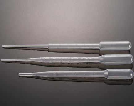
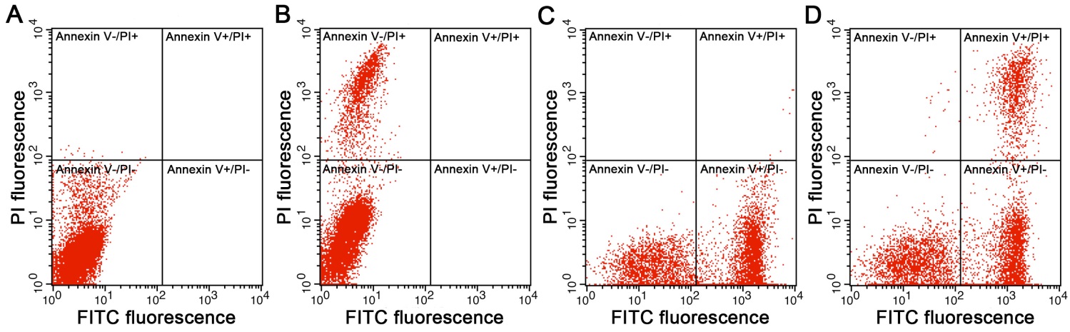
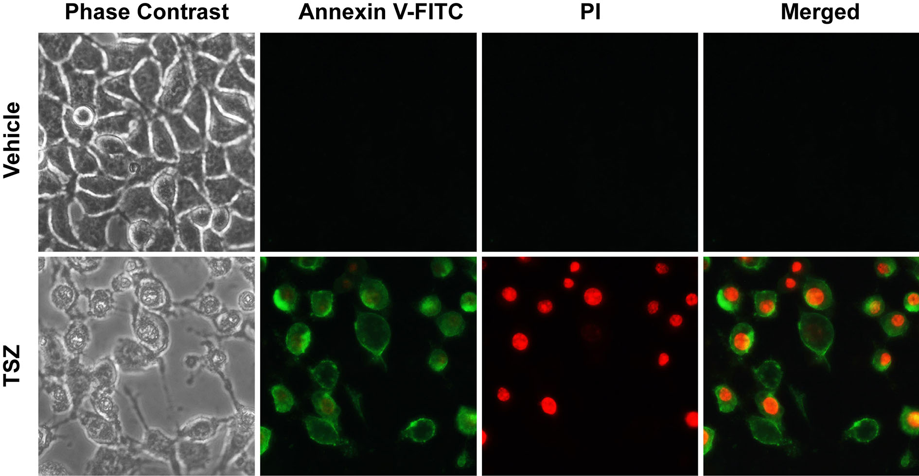
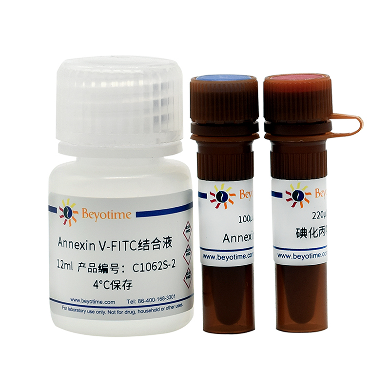
使用说明: 1. 对于悬浮细胞: a. 在进行完细胞凋亡刺激后,1000g离心5分钟,弃上清,收集细胞,用PBS轻轻重悬细胞并计数。注意:PBS重悬不能省略,PBS重悬的过程同时也起到了洗涤细胞的作用,可以保证后续Annexin V-FITC的结合。 b. 取5-10万重悬的细胞,1000g离心5分钟,弃上清,加入195μl Annexin V-FITC结合液轻轻重悬细胞。 c. 加入5μl Annexin V-FITC,轻轻混匀。 d. 加入10μl碘化丙啶染色液,轻轻混匀。 e. 室温(20-25℃)避光孵育10-20分钟,随后置于冰浴中。可以使用铝箔进行避光。孵育过程中可以重悬细胞2-3次以改善染色效果。 f. 如果用于流式细胞仪检测,可立即上机检测,Annexin V-FITC为绿色荧光,碘化丙啶(PI)为红色荧光,流式检测细胞凋亡的效 果及其验证请参考图1和图2。初次进行流式细胞仪检测时,建议选择一组适当的细胞参考图2设置未染色、PI单染和Annexin V-FITC单染这3个对照。如果用于荧光显微镜检测,1000g离心5分钟,收集细胞,用50-100μl Annexin V-FITC结合液轻轻重悬细胞,涂片后,荧光显微镜下观察。注意:细胞在染色后须尽快完成检测,通常宜在1小时之内完成检测。用于流式细胞仪检测时,如果发现Annexin V-FITC单独染色时出现了过多的PI假阳性细胞,并且通过调整相关设置和参数也无法改善,可以用PBS将Annexin V-FITC稀释3-10倍后再进行检测。 2. 对于贴壁细胞的消化后检测: a. 把细胞培养液吸出至一合适离心管内,PBS洗涤贴壁细胞一次,加入适量胰酶细胞消化液(可含有EDTA)消化细胞。室温孵育至轻轻吹打可以使贴壁细胞吹打下来时,吸除胰酶细胞消化液。需避免胰酶的过度消化。注意:对于贴壁细胞,胰酶消化步骤很关键。胰酶消化时间如果过短,细胞需要用力吹打才能脱落,容易造成细胞膜的损伤,从而导致细胞坏死的假阳性;消化时间如果过长,同样易造成细胞膜损伤而出现细胞坏死的假阳性,甚至会影响细胞膜上磷脂酰丝氨酸与Annexin V-FITC的结合从而干扰对于细胞凋亡的检测。同时,胰酶细胞消化液中应尽量不含EDTA,因为EDTA可能会影响Annexin V与磷脂酰丝氨酸的结合。 b. 加入步骤2a中收集的细胞培养液,把细胞轻轻吹打下来,转移到离心管内,1000g离心5分钟,弃上清,收集细胞,用PBS轻轻重悬细胞并计数。注意:加入步骤2a中的细胞培养液非常重要,一方面可以收集已经悬浮的发生凋亡或坏死的细胞,另一方面细胞培养液中的血清可以有效抑制或中和残留的胰酶。残留的胰酶会消化并降解后续加入的Annexin V-FITC,导致染色失败。 c. 取5-10万重悬的细胞,1000g离心5分钟,弃上清,加入195μl Annexin V-FITC结合液轻轻重悬细胞。 d. 加入5μl Annexin V-FITC,轻轻混匀。 e. 加入10μl碘化丙啶染色液,轻轻混匀。 f. 室温(20-25℃)避光孵育10-20分钟,随后置于冰浴中。可以使用铝箔进行避光。孵育过程中可以重悬细胞2-3次以改善染色效果。 g. 如果用于流式细胞仪检测,可立即上机检测,Annexin V-FITC为绿色荧光,碘化丙啶(PI)为红色荧光,流式检测的效果及其验 证请参考图1和图2。如果用于荧光显微镜检测,1000g离心5分钟,收集细胞,用50-100μl Annexin V-FITC结合液轻轻重悬细胞,涂片后,荧光显微镜下观察。注意:细胞在染色后须尽快完成检测,通常宜在1小时之内完成检测。用于流式细胞仪检测时,如果发现Annexin V-FITC单独染色时出现了过多的PI假阳性细胞,并且通过调整相关设置和参数也无法改善,可以用PBS 将Annexin V-FITC稀释3-10倍后再进行检测。 3. 对于贴壁细胞的原位荧光检测: 注:本方法的优点是可以原位观察细胞凋亡,缺点是部分凋亡由于不贴壁而检测不到。 a. (选做)如果条件许可,把细胞培养于24孔板、48孔板或96孔板内。在凋亡诱导结束后,用可以对多孔板进行离心的离心机1000g离心5分钟。 b. 吸除细胞培养液,加入PBS洗涤一次。如果条件许可,在吸除PBS前1000g离心5分钟。 c. 加入195μl Annexin V-FITC结合液。 d. 加入5μl Annexin V-FITC,轻轻混匀。 e. 加入10μl碘化丙啶染色液,轻轻混匀。 f. 室温(20-25℃)避光孵育10-20分钟,随后置于冰浴中。可以使用铝箔进行避光。 g. 随即在荧光显微镜下观察,Annexin V-FITC为绿色荧光,碘化丙啶(PI)为红色荧光。注意:细胞在染色后须尽快完成检测,通常宜在1小时之内完成检测。
Effect of Bufotalin on the Apoptosis of BGC-823 Cells.
中药与新药临床药理学 .2007 Mar;18(2):117-9.
2. Yang L, Zhou X, Yang J, Yin X, Han L, Zhao D.
Aspirin inhibits cytotoxicity of prion peptide PrP106-126 to neuronal cells associated with microglia activation in vitro.
J Neuroimmunol . 2008 Aug 13;199(1-2):10-7. (IF 3.125)
3. Gao C, Jiang Y, Tan C, Zu X, Liu H, Cao D.
Synthesis and potent antileukemic activities of 10-benzyl-9(10H)-acridinones.
BIOORG MED CHEM LETT . 2008 Sep 15;16(18):8670-5. (IF 2.572)
4. Wang LM, Li QY, Zu YG, Fu YJ, Chen LY, Lv HY, Yao LP, Jiang SG.
Anti-proliferative and pro-apoptotic effect of CPT13, a novel camptothecin analog, on human colon cancer HCT8 cell line.
CHEM-BIOL INTERACT . 2008 Nov 25;176(2-3):165-72. (IF 3.723)
5. Wang L, Li Z, Wang C, Yang Y, Sun L, Yao W, Cai X, Wu G, Zhou F, Zha X.
E-cadherin decreased human breast cancer cells sensitivity to staurosporine by up-regulating Bcl-2 expression.
Arch Biochem Biophys . 2009 Jan 1;481(1):116-22. (IF 3.391)
6. Yan HL, Xue G, Mei Q, Wang YZ, Ding FX, Liu MF, Lu MH, Tang Y, Yu HY, Sun SH.
Repression of the miR-17-92 cluster by p53 has an important function in hypoxia-induced apoptosis.
EMBO J . 2009 Sep 16;28(18):2719-32. (IF 9.889)
7. Chen YJ, Huang XB, Li ZX, Yin LL, Chen WQ, Li L.
Tenuigenin protects cultured hippocampal neurons against methylglyoxal-induced neurotoxicity.
Eur J Pharmacol . 2010;645(1-3):1-8. (IF 3.263)
8. Wang P, Xu CS, Xu J, Wang X, Leung AW.
Hypocrellin B enhances ultrasound-induced cell death of nasopharyngeal carcinoma cells.
Ultrasound Med Biol . 2010;36(2):336-42. (IF 2.514)
9. Luo M, Liu X, Zu Y, Fu Y, Zhang S, Yao L, Efferth T.
Cajanol, a novel anticancer agent from Pigeonpea [Cajanus cajan (L.) Millsp.] roots, induces apoptosis in human breast cancer cells through a ROS-mediated mitochondrial pathway.
10. Shao D, Zhong X, Zhou YF, Han Z, Lin Y, Wang Z, Bu L, Zhang L, Su XD, Wang H.
Structural and functional comparison of MIF ortholog from Plasmodium yoelii with MIF from its rodent host.
Mol Immunol . 2010;47(4):726-37. (IF 3.641)
11. Wang X, Xu J, Ju S, Ni H, Zhu J, Wang H.
Livin gene plays a role in drug resistance of colon cancer cells.
Clin Biochem . 2010;43(7-8):655-60. (IF 2.573)
12. Du S, Zhou J, Jia Y, Huang K.
SelK is a novel ER stress-regulated protein and protects HepG2 cells from ER stress agent-induced apoptosis.
Arch Biochem Biophys . 2010 Oct 15;502(2):137-43. (IF 3.391)
13. Zhang J, Tang Y, Li S, Liao C, Guo X.
Targeting of the B-lineage leukemia stem cells and their progeny with norcantharidin encapsulatedliposomes modified with a novel CD19 monoclonal antibody 2E8 in vitro.
J Drug Target . 2010 Nov;18(9):675-87. (IF 3.38)
14. Rasul A, Khan M, Yu B, Ma T, Yang H.
Xanthoxyletin, a coumarin induces S phase arrest and apoptosis in human gastric adenocarcinomaSGC-7901 cells.
ASIAN PAC J CANCER P . 2011;12(5):1219-23. (IF 1.24)
15. Wang CM, Sheng GY, Lu J, Xie L, Bai ST, Xu XJ, Liu YF.
Effect of small interfering RNA targeting wild-type FLT3 in acute myeloid leukaemia cells in vitroand in vivo.
J Int Med Res . 2011;39(5):1661-74. (IF 1.287)
16. Zhong MR, Zhao YH, Zhang K, Yang LF, Zheng YC, He CY.
Inhibitory Effects of Natural Compound Alantolactone on Human Non-small Cell Lung Cancer A549 Cells.
CHEM RES CHINESE U . 2011;27(2):241-4. (IF 1.063)
17. Ji YB, Qu ZY, Zou X.
Juglone-induced apoptosis in human gastric cancer SGC-7901 cells via the mitochondrial pathway.
1,3,4-tri-O-galloyl-6-O-caffeoyl-β-D-glucopyranose, a new anti-proliferative ellagitannin,regulatesthe expression of microRNAs in HepG(2) cancer cells.
南方医科大学学报 . 2011 Oct;31(10):1641-8.
36. Han JB, Tao ZZ, Chen SM, Kong YG, Xiao BK.
Adenovirus-mediated transfer of tris-shRNAs induced apoptosis of nasopharyngeal carcinoma cellin vitro and in vivo.
Cancer Lett . 2011 Oct 28;309(2):162-9. (IF 7.36)
37. Wang Z, Yang X, Yang S, Ren G, Ferreri M, Su Y, Chen L, Han B.
Sodium fluoride suppress proliferation and induce apoptosis through decreased insulin-likegrowth factor-I expression and oxidative stress in primary culturedmouse osteoblasts.
38. Yang YF, Chen Z, Hu SL, Hu J, Li B, Li JT, Wei LJ, Qian ZM, Lin JK, Feng H, Zhu G.
Interleukin-1 receptor associated kinases-1/4 inhibition protects against acute hypoxia/ischemia-induced neuronal injury in vivo and in vitro.
Neuroscience . 2011 Nov 24;196:25-34. (IF 3.056)
39. Tu ZZ,Li H,Ma YX,Tang B,Tian JM,Akers W,Achilefu S and Gu YQ.
The enhanced antiproliferative response to combined treatment of trichostatin A with raloxifene in MCF-7 breast cancer cells and its relevance to estrogen receptor β expression.
Molecular and Cellular Biochemistry .2012;366:111-22. (IF 2.884)
40. SU JQ,CHI BR,LI X,LIU L,LIU LM,QI YX,WANG ZY,JIN NY.
Inhibition of Dual Specific Oncolytic Adenovirus on Esophageal Cancer via Activation of Caspases by a Mitochondrial-dependent Pathway.
高等学校化学研究:英文版 CAS CSCD.2012;28(3):465-71.
41. Liu X, Zhao M, Lu J, Ma J, Wei J, Wei S.
Cell responses to two kinds of nanohydroxyapatite with different sizes and crystallinities.
INT J NANOMED . 2012;7:1239-50. (IF 5.115)
42. Gu S, He J.
Daphnoretin induces cell cycle arrest and apoptosis in human osteosarcoma (HOS) cells.
Molecules . 2012 Jan 9;17(1):598-612. (IF 3.267)
43. Shi G, Zhang Z, Zhang R, Zhang X, Lu Y, Yang J, Zhang D, Zhang Z, Li X, Ning G.
Protective effect of andrographolide against concanavalin A-induced liver injury.
57. Zou X, Feng Z, Li Y, Wang Y, Wertz K, Weber P, Fu Y, Liu J.
Stimulation of GSH synthesis to prevent oxidative stress-induced apoptosis by hydroxytyrosol inhuman retinal pigment epithelial cells: activation of Nrf2 and JNK-p62/SQSTM1 pathways.
The Total Starch (AA/AMG) test kit is used for the measurement and analysis of total starch in cereal flours and food products. This kit now contains an improved α-amylase that allows the amylase incubations to be performed at pH 5.0 (as well as pH 7.0).
Colourimetric method for the determination of Total Starch in
cereal products, feeds, foodstuffs and other materials
The pH of the assay solution after the sample is added should be the same as that of the assay buffer that is supplied with the kit.
Low sample volumes (e.g. 0.1 mL) are not likely to affect the pH of the assay solution and therefore may not require pH adjustment.
Samples above 0.1 mL are more likely to affect the pH of the assay solution and therefore the pH of these samples should be adjusted as described in the data booklet, prior to addition to the assay.
The Regular Maize Starch does not require DMSO pre-treatment. The value should be about 84% with a moisture content of about 12%, the final dry weight value is about 96-97%. Store the sample at room temperature, dry.
If you suspect that the Megazyme test kit is not performing as expected such that expected results are not obtained please do the following:
Ensure that you have tested the standard sample that is supplied with the Megazyme test kit.
Send the results of the kit standard, blank samples and the results obtained for your sample, in the relevant MegaCalc spreadsheet (if available) to Megazyme (cs@megazyme.com). Where available the relevant MegaCalc spreadsheet can be downloaded from where the product appears on the Megazyme website.
State the kit lot number being used (this is found on the outside of the kit box).
State which assay format was used (refer to the relevant page in the kit booklet if necessary).
State exact details of any modifications to the standard procedure that is provided by Megazyme.
State the sample type and describe the sample preparation steps if applicable.
Yes. We believe that the DMSO step will solubilise vitrified starch in malt. Make sure that the malt is milled to pass a 0.5 mm screen. You could vary the time of cooking with DMSO to check solubilisation (i.e. 5 minutes, 10 minutes, or even up to 1 hour).
There should be no problem in measuring the starch in plasterboard. I suggest that you grind about 100 g in a kitchen blender and then fine mill to pass 0.5 mm screen. Run a standard assay, but adjust volume to 10 mL after alpha-amylase treatment. Keep a close check on the pH. Plasterboard may push the pH value up (pH up to about 8 should be fine). You may be advised to run a DMSO format concurrently just to be sure. When you treat with amyloglucosidase, I would advise that you take 0.2 and 0.4 mL aliquots of digest (to get the colour up), also, be careful about checking the pH.
Our Total Starch Assay Kit could measure starch left in a residue, or starch extracted. No method could measure potential extractable starch, as this will depend on numerous factors, including processing equipment, conditions etc.
Most of the maltodextrins can be removed with 50% ethanol washing. If the starch is not gelatinised, it can be washed with cold water. This will remove all of the soluble maltodextrins, but the starch will spin down. If the starch has been gelatinised, then the best material which can be used for washing is 50% ethanol.
We think that calcium carbonate etc. will not cause any problems. However, this of course depends on the amount present and if it changes the pH of the incubation mixture.
The method will work for some chemically modified starches (e.g. crosslinked) however, if the degree of chemical modification is high, there will be an underestimation as the modification will interfere with complete hydrolysis to glucose and subsequent measurement.
You only need to wash samples which you feel may contain glucose and/or maltodextrins, e.g. breakfast cereals. There is little glucose in ground cereals, so it is not necessary to pre-wash these materials.
The Starch Damage Kit may be best for this. If the starch is gelatinised and dried before analysis the correct results for gelatinisation will not be obtained.
DMSO does solubilise resistant starch (crystallise amylose and amylopectin). The only starch material we have had problems in dissolving in DMSO is potato amylose.
We believe that for starch fragments, oligosaccharides of a DP up to 10 would be soluble in 80% alcohol. The degree of solubility of other oligosaccharides would depend on the sugar type and linkage type.
The Total Starch Kit can be used for liquids containing as little as 200 micrograms per mL with some adjustments of conditions, as below:
Mix 0.5 mL of sample with 0.5 mL of 100 mM sodium acetate buffer (pH 4.5). Incubate at 40˚C and add 0.1 mL of Amyloglucosidase and incubate for 30 minutes. Add GOPOD reagent as usual. You will need to run an AMG blank as this enzyme preparation contains a very small amount of glucose.
The AMG activity was determined with soluble starch as substrate (10 mg/mL) in 0.1 M sodium acetate buffer at pH 4.5 and 40˚C. One Unit is the amount of enzyme required to hydrolyse one micromole of maltose per minute (i.e. to release 2 micromoles of glucose). Glucose release is measured with Glucose Determination Reagent.
We can assure you that the Total Starch Kit will not work if incubations with the thermostable alpha-amylase are performed at pH 3 or 4. This enzyme is inactivated at pH values below 5.0. You may wish to look at a method using just amyloglucosidase which is quite active down to pH 4.0. Check the old AOAC procedure for starch.
When we dispense the enzymes we usually include an extra 5% in each vial, so yes, you can dilute the whole vial. When you do this, please divide into aliquots and store them frozen.
You can use the standard procedure. We would recommend that you treat 2 mL of beer or wort with 8 mL of ethanol, stir and centrifuge (3,000 rpm). Wash the pellet with 10 mL of 80% ethanol. Then dissolve/suspend the pellet in 2 mL of sodium acetate buffer (pH 4.5, 0.1 M) and cook at 100˚C for 10 min. Then add 0.1 mL AMG from the Total Starch Kit and proceed according to the method. You will have to determine the degree of dilution for yourself. Treat 0.1 mL with GOPOD etc.